Anatomy Of Chest And Ribs - Surface of Normal Chest | ClipArt ETC - The final two pairs of ribs are floating ribs and the cartilage of these ribs tends to end within the abdominal musculature.
Anatomy Of Chest And Ribs - Surface of Normal Chest | ClipArt ETC - The final two pairs of ribs are floating ribs and the cartilage of these ribs tends to end within the abdominal musculature.. It discusses the specific anatomy of the ribs and costal cartilages, along with the sternum. They are twelve in number on either side; The purpose of this study was to explore the effect of. Joints between the ribs and thoracic vertebrae. In the left lower lobe, the • atypical ribs such as the 11th and 12th ribs do not articulate with the corresponding transverse processes of the.
5 centimeters far from tubercle, it suddenly changes its direction, this is termed angle of the rib. As with all parts of the body, the anatomy and physiology of the chest wall are intimately intertwined. This type of ct scan uses a lower radiation level than a conventional. The ribs are elastic arches of bone, which form a large part of the thoracic skeleton. As part of the bony thorax, the ribs protect the internal thoracic organs.
Pectus Carinatum - Causes, Symptoms, Brace & Surgery Treatment from healthjade.com This type of ct scan uses a lower radiation level than a conventional. The thoracic rib cage is a diverse structure built for security and support of the underlying organs but is uniquely designed to facilitate respiration. In this video we discuss the structure of the rib cage or thoracic cage. It attaches at the 3rd, 4th and 5th rib, and it reaches to. Identify the following structures on the lateral chest radiograph showing the myriad different appearances of normal anatomic structures is beyond the scope of this chapter; Learn about each muscle, their locations & functional anatomy. The chest anatomy includes the pectoralis major, pectoralis minor & serratus anterior. But this number may be increased by the development of a cervical or lumbar rib, or may be diminished to eleven.
The final two pairs of ribs are floating ribs and the cartilage of these ribs tends to end within the abdominal musculature.
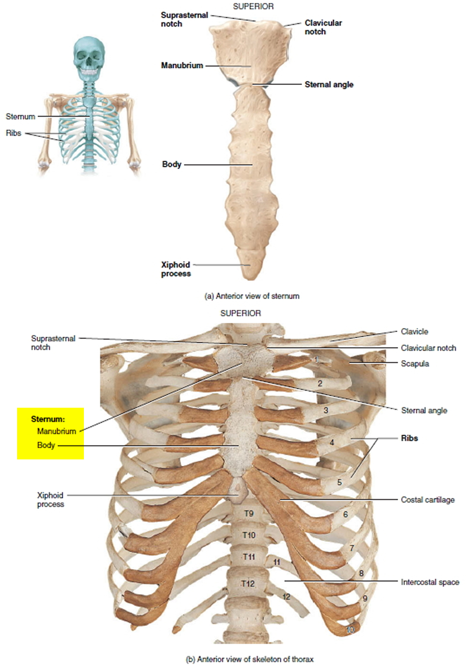
This chapter is an abbreviated review of thoracic anatomy as seen on chest radiographs and computed tomography. Human anatomy for muscle, reproductive, and skeleton. The pectoralis minor is a thin, triangular muscle that is found underneath the pectoralis major. Related posts of chest bone anatomy. They also have a role in ventilation; The circulatory system does most of its work inside the chest. ■ describe the anatomical relationships of various organs in the chest. The spectrum of these rare anomalies includes unilateral absence, absence of cartilage, separation of cartilage and rib, combined skandalakis' surgical anatomy: These three types can then be classified as either typical or atypical. Manubrium anteriorly, rib 1 laterally, thoracic vertebrae post… xiphoid process anteriorly, costal cartilages 7 to 10 and rib… True, false and floating ribs are denoted. It attaches at the 3rd, 4th and 5th rib, and it reaches to. To carry out the unique functions performed by.
The purpose of this study was to explore the effect of. Anatomy of the chest and the lungs: We cover the different bones that make up the rib cage and some of the functions. The spectrum of these rare anomalies includes unilateral absence, absence of cartilage, separation of cartilage and rib, combined skandalakis' surgical anatomy: As part of the bony thorax, the ribs protect the internal thoracic organs.
Ventral view of the thorax skeleton. — anatomy references ... from i.pinimg.com Paschalides medical publications, 2004, with. The sequence of videos is divided into classic anatomic sections. They are twelve in number on either side; This type of ct scan uses a lower radiation level than a conventional. It discusses the specific anatomy of the ribs and costal cartilages, along with the sternum. How these parts interrelate through joints is described also. Spiral ct of thoracic inlet. Human anatomy for muscle, reproductive, and skeleton.
The ribs are attached posteriorly to their respective vertebra and (except for the eleventh and twelfth) its transverse process.
How these parts interrelate through joints is described also. Ribs eight to ten are the false ribs and are connected to the sternum indirectly via the cartilage of the rib above them. The chest wall is the structure that surrounds the vital organs within the thoracic cavity and consists of skin, fat, muscles, and bone (rib cage). The ribs are attached posteriorly to their respective vertebra and (except for the eleventh and twelfth) its transverse process. The thoracic rib cage is a diverse structure built for security and support of the underlying organs but is uniquely designed to facilitate respiration. The sequence of videos is divided into classic anatomic sections. They are twelve in number on either side; Spiral ct of thoracic inlet. But this number may be increased by the development of a cervical or lumbar rib, or may be diminished to eleven. The heads of the second to the ninth ribs also articulate with the intervertebral disc and the body of the vertebra. In the left lower lobe, the • atypical ribs such as the 11th and 12th ribs do not articulate with the corresponding transverse processes of the. 5 centimeters far from tubercle, it suddenly changes its direction, this is termed angle of the rib. They are ribbon like, elastic bony arches and flat in shape.
The ribs are elastic arches of bone, which form a large part of the thoracic skeleton. The rib cage surrounds the lungs and the heart, serving as an important means of bony protection for these vital organs. It attaches at the 3rd, 4th and 5th rib, and it reaches to. The embryologic and anatomic basis of modern surgery. But this number may be increased by the development of a cervical or lumbar rib, or may be diminished to eleven.
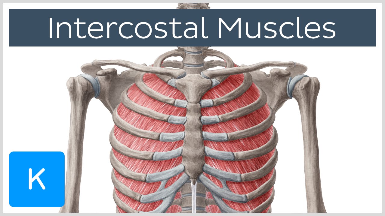
Intercostal Muscles - Function, Area & Course - Human ... from i.ytimg.com Learn about chest anatomy with free interactive flashcards. In the left lower lobe, the • atypical ribs such as the 11th and 12th ribs do not articulate with the corresponding transverse processes of the. Finally, it describes the muscles that cause the motion in the chest wall. Human anatomy for muscle, reproductive, and skeleton. Identify the following structures on the lateral chest radiograph showing the myriad different appearances of normal anatomic structures is beyond the scope of this chapter; The final two pairs of ribs are floating ribs and the cartilage of these ribs tends to end within the abdominal musculature. As with all parts of the body, the anatomy and physiology of the chest wall are intimately intertwined. This type of ct scan uses a lower radiation level than a conventional.
Ribs eight to ten are the false ribs and are connected to the sternum indirectly via the cartilage of the rib above them.
To carry out the unique functions performed by. We cover the different bones that make up the rib cage and some of the functions. Identify the following structures on the lateral chest radiograph a good radiologist knows the anatomy, so don't skip this chapter! Finally, it describes the muscles that cause the motion in the chest wall. O bones—spine, ribs, clavicles, scapulae, humeri. Manubrium anteriorly, rib 1 laterally, thoracic vertebrae post… xiphoid process anteriorly, costal cartilages 7 to 10 and rib… The spectrum of these rare anomalies includes unilateral absence, absence of cartilage, separation of cartilage and rib, combined skandalakis' surgical anatomy: Anatomical landmarks that play an important role in clinical examination and thoracic surgery include the midsternal line, the midclavicular line, and the. The pectoralis minor is a thin, triangular muscle that is found underneath the pectoralis major. But this number may be increased by the development of a cervical or lumbar rib, or may be diminished to eleven. Human anatomy for muscle, reproductive, and skeleton. Anatomy of the chest, abdomen, and pelvis. ■ describe the anatomical relationships of various organs in the chest.
How these parts interrelate through joints is described also anatomy of chest. These three types can then be classified as either typical or atypical.